Electron microscope image processing and analysis are segmented into two main areas: general image processing (IPA) and crystallographic image processing (CIP). Central to these processes is the Fast Fourier Transform (FFT) technique. In EMIPA (Electron Microscope Image Processing and Analysis): the experimental image can be resized and rotated image. Part of the image can be selected and saved with a new scale bar. A series of linear profiles can be retrieved. Typical filters are available for FFT and IFFT processing. The indices can be added for the FFT pattern with an auxiliary tool. Histogram of the IFFT image can be optimized. Some important features are listed below,
Here is an example of a TEM study on crystal structures in the rare-earth-free intermetallic alloys, Fe3+xCo3-xTi2 (x = 0, 1, 2, 3). In these alloys, the main compound belongs to Laves C14 variant surrounded by α-Fe type crystal as the secondary phase in the Fe3+xCo3-xTi2 (x = 0, 1, 2, 3) alloys, in agreement with the Fe-Ti and Fe-Co-Ti phase diagrams. |
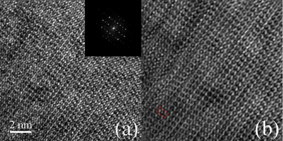
(a) HREM image and (b) the IFFT processing image of the main compound in the Fe3Co3Ti2 alloy.