
EMCIP (Electron Microscope Crystallographic Image Processing) adopts crystallographic image processing (CIP), a technique to enhance the HREM image for the structure determination. The principles are based that crystallographic structure factor phase information is present HREM images and can be utilized for structure analysis. EMCIP has been developed for the enhancing the experimental image with the CIP technique. A contrast transfer function is included for correcting the crystallographic phase in the FFT data. Experimental electron diffraction intensities can be used to replace diffraction intensities in the FFT data. The image can be processed using pre-built the17 plane symmetry groups and displayed in the pseudo-color image and contour map. Some important features are listed below,
|
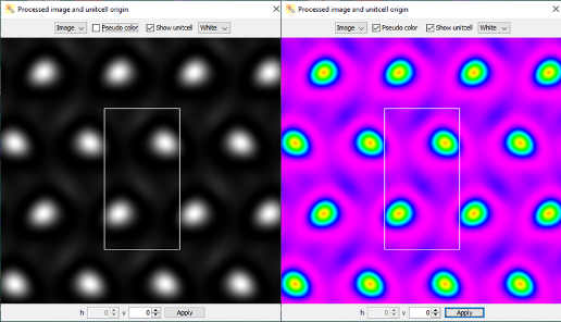
An example of the CIP image in gray scale and in pseudo color after image crystallography processing.